Figure 4.
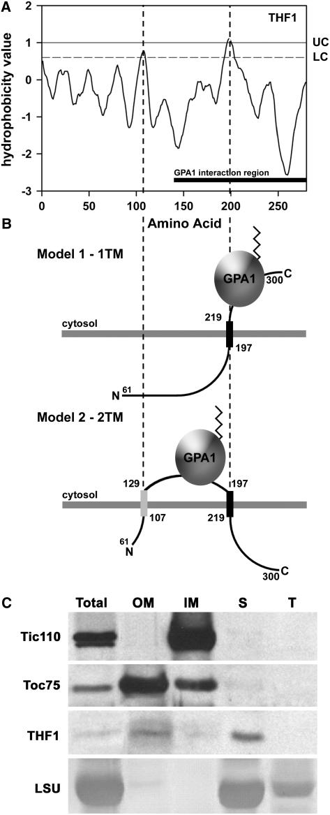
THF1 Is a Protein of the Outer Plastid Membrane and Stroma.
(A) Kyte–Doolittle hydropathy plot for THF1. UC and LC denote the upper and lower cutoffs for the prediction of membrane-spanning domains, respectively. The solid bar at bottom denotes the positions mapped by the yeast two-hybrid assay as the THF1–GPA1 interaction domain. Dashed lines denote positions of the two predicted membrane spans.
(B) Possible topologies for THF1 that enable interaction between the membrane-spanning pool of THF1 and the plasma membrane–delimited GPA1. THF1 contains at least one (model 1), and possibly two (model 2), membrane-spanning domains, which embed THF1 in plastid membranes. The angled line on GPA1 represents the N-terminal myristoyl group that anchors GPA1 to the plasma membrane (data not shown).
(C) Isolated plastids were fractionated and subjected to immunoblot analysis with the corresponding antisera to the indicated marker proteins or THF1. Fractions are represented as follows: Total, whole preparation; OM, outer plastid membrane; IM, inner plastid membrane; S, stromal fraction; T, thylakoid fraction. Controls used to determine THF1 localization were as follows: Tic110, a component of the inner plastid membrane; Toc75, a component of the outer plastid membrane; and the large subunit of ribulose-1,5-bisphosphate carboxylase/oxygenase (LSU), which is found in the stroma and thylakoid.

