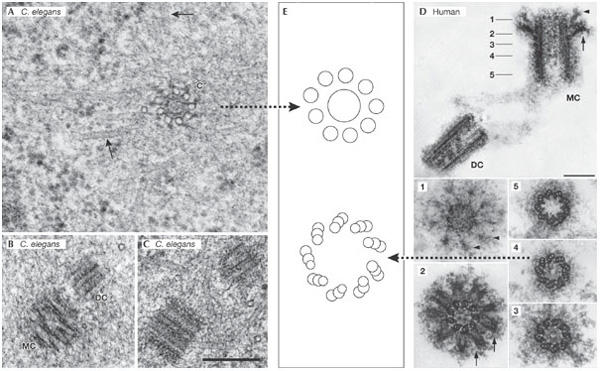
Figure 2.

Human and worm centriolar structure: similar design, different complexity. (A–C) Centrioles of the Caenorhabditis elegans embryo. Scale bar, 250 nm. (A) Transmission electron micrograph of a prometaphase centriole (c) with singlet microtubules in cross section. Spindle microtubules are indicated by arrows. (B) Mother centriole (MC) and daughter centriole (DC) in prometaphase. (C) A centriole pair in interphase. (D) A human centrosome isolated from the human KE37 lymphoblastic cell line that was used for the proteome analysis by Andersen et al (2003). The five numbered panels below correspond to sections through the MC at the points indicated in the upper panel. The arrowheads and the arrows identify the distal and sub-distal appendages, respectively. Scale bars, 0.2 mm. (E) Cartoons depicting the microtubule architecture of the indicated centrioles. (A–C) were generously provided by T. Müller-Reichert (Dresden, Germany) and (D) by M. Bornens (Paris, France).
