
Figure 3.
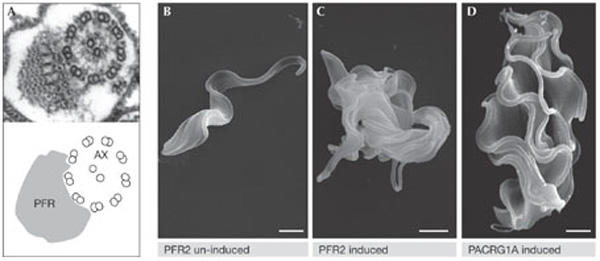
Disruption of flagellar structure blocks cytokinesis of Trypanosomas brucei. (A) A transmission electron micrograph showing the 9 + 2 structure of the axoneme (AX) alongside the paraflagellar rod (PFR). (B–D) Scanning electron micrographs showing bloodstream-form T. brucei before (B) and after ablation of the flagellar proteins PFR2 (C) and PACRG1A (D). Images were kindly provided by K. Gull (Oxford, UK).