
Figure 4.
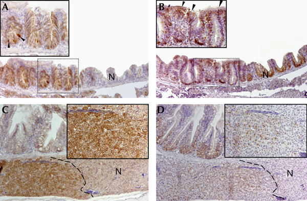
Intestinal and hepatic adenomas in 7,12-dimethylbenz[a]anthracene-treated apc/+ fish. (A) Section of the intestine of a 6-month-old 7,12-dimethylbenz[a]anthracene (DMBA)-treated apc/+ fish stained with β-catenin. A region with high levels of β-catenin compared with the adjacent normal tissue (N) is observed. The inset depicts a high-magnification image of the boxed area of the adenoma in which cells have accumulated high levels of cytoplasmic and nuclear β-catenin (arrowheads). (B) Serial section of the adenoma shown in (A) stained for proliferating-cell nuclear antigen (PCNA). Note that most of the cells in the structure are proliferating. The inset depicts a higher magnification image of the boxed area of the adenoma. Ectopic PCNA+ cells are found in the surface epithelium (arrowheads). (C) Section of liver and small intestine of a 6-month-old DMBA-treated apc/+ fish stained for β-catenin. A confined liver lesion demarcated by a striped line with high levels of β-catenin is depicted. The inset shows a high-magnification image of hepatocytes that have accumulated nuclear and cytoplasmic β-catenin. (D) Serial section of the lesion shown in (C) stained for PCNA. Cells in the lesion (demarcated by a striped line) are proliferating. The inset depicts a higher magnification image of PCNA+ cells. Original magnification: (A–D) × 100; insets × 200.