
Figure 1.
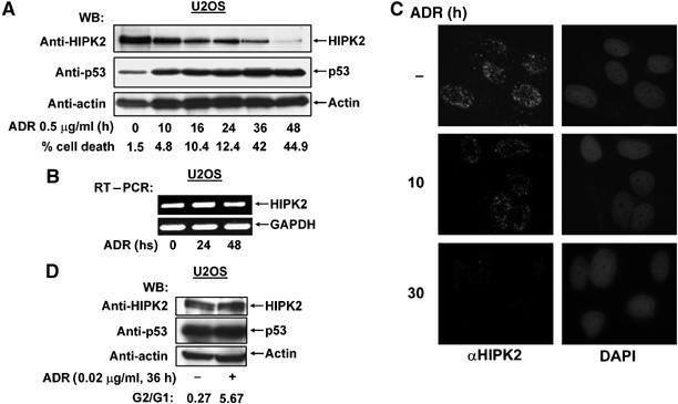
DNA damage triggers HIPK2 degradation. (A) U2OS cells were treated for the indicated periods with adriamycin (ADR). Equal amounts of proteins contained in cell extracts were separated by SDS–PAGE and analyzed by Western blotting (WB) for the occurrence of endogenous HIPK2, p53 and the loading control β-actin as shown. Apoptosis in these cells was quantified by FACS analysis, typical values are shown. (B) U2OS cells were treated with adriamycin (0.5 μg/ml) as shown. Relative mRNA levels of HIPK2 and the control GAPDH were determined by RT–PCR with specific primers, an ethidium bromide-stained agarose gel is displayed. (C) U2OS cells were treated with adriamycin (0.5 μg/ml) as shown and the localization of endogenous HIPK2 was revealed by indirect immunefluorescence. Nuclear DNA was stained with DAPI. (D) U2OS cells remained untreated or received 0.02 μg/ml adriamycin for 36 h. Cells were analyzed by FACS analysis for cell cycle distribution and by Western blotting for the occurrence of HIPK2, p53 and β-actin.