
Figure 2.
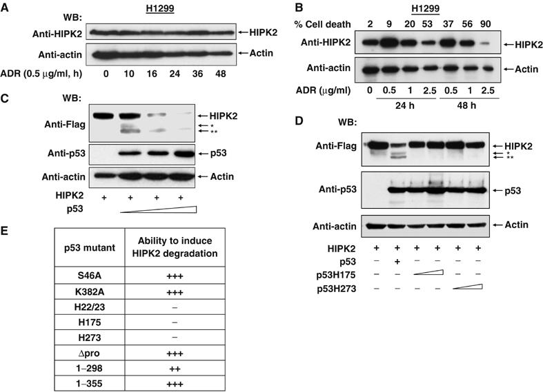
p53 activation triggers HIPK2 degradation. (A) p53-deficient H1299 cells were treated and analyzed as in (Figure 1A). (B) H1299 cells were treated with increasing concentrations of adriamycin for 24 or 48 h as shown. Cells were analyzed for apoptosis and by Western blotting for HIPK2 and the loading control β-actin. Note that cells showing massive apoptosis also contain less of the loading control. (C) H1299 cells were transiently transfected to express a constant amount of Flag-tagged HIPK2 and increasing concentrations of p53. Cell extracts were prepared and the occurrence of HIPK2 and p53 was detected by immunoblotting. The positions of HIPK2* and HIPK2** cleavage products are indicated. (D) H1299 cells were transfected to express Flag-HIPK2 in the absence or presence of p53 or escalating doses of the indicated p53 point mutants. After 24 h, cell extracts were prepared and analyzed by Western blotting. (E) Summary of the experiments measuring the impact of p53 mutants on their ability to trigger HIPK2 processing (+++ complete cleavage, ++ cleavage less efficient, − no cleavage).