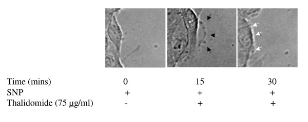
Figure 4.
Thalidomide attenuates NO mediated migration in ECs: After the wound was created in confluent ECs, they were treated with 500μM SNP (NO donor) and incubated for 15 minutes at 37°C/5% CO2. Next, the cells were treated with 75 μg/ml thalidomide and incubated for another 15 minutes. Images of the wound-edge were taken at 0 minutes, 15 minutes and 30 minutes with a 40× magnification objective lens mounted on Nikon TE2000-U inverted microscope. Arrows indicate the growing phases of the wounds after 15 minutes of incubation with SNP. Arrows in the middle panel indicates the growing phase of ECs under thalidomide treatment, while thalidomide arrests the SNP mediated migration of the cells after 15 minutes of thalidomide treatments (white arrows in the right panel).

