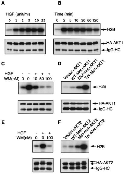
Figure 1.
Activation of AKT1 and AKT2 by HGF or active forms of Met. A human HA-AKT1 expression plasmid was transfected into SK-LMS-1 cells. Cells were serum starved for 16 h, followed by stimulation at increasing concentrations of HGF for 10 min (A) or with 10 units/ml HGF for various times (B). (C) AKT1 activity is stimulated by 10 units/ml HGF and inhibited by WM. (D) SK-LMS-1 cells were transiently cotransfected with an HA-AKT1 plasmid together with pMB1 vector, WT-Met, Met-mut, or Tpr-Met. AKT1 is strongly activated by Met-mut and Tpr-Met. (E) AKT2 activity is also stimulated by HGF and inhibited by WM. (F) AKT2 is activated by Met-mut and Tpr-Met. (A–F Upper) Akt kinase activity. Expressed HA-AKT1 or HA-AKT2 was immunoprecipitated with an anti-HA antibody, and kinase assays were performed as described in Materials and Methods. IgG-HC, immunoglobulin heavy chain. (A–F Lower) Western blot analysis of immunoprecipitates by using anti-HA antibody, demonstrating equivalent loading in each lane. Slower mobility bands in E and F are presumed to be the fully phosphorylated activated form of HA-AKT2 (17).

