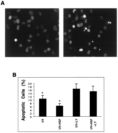
Figure 2.
Anti-apoptotic signaling by HGF in NIH 3T3 cells transfected with WT-Met. (A) Cells were serum starved overnight, and after treatment with UV irradiation, fresh serum-free medium, supplemented with (Left) or without (Right) HGF, was added to the cells. Cells were incubated for another 24 h and harvested for evaluation of apoptosis by Hoechst 33342 staining. Normal nuclei show faint delicate chromatin staining, nuclei at the early stage of apoptosis display increased condensation and brightness, and nuclei at the late stage of apoptosis exhibit chromatin condensation and nuclear fragmentation. (B) Apoptotic cells with characteristic chromatin condensation and nuclear fragmentation were counted and expressed as a percentage of the total cell number. Bar = mean ± SD of three independent experiments. *, P < 0.01.

