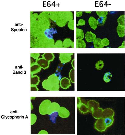
Figure 2.
Localization of erythrocytic proteins and merozoites in schizont-containing blood smears cultured in the presence or absence of E64. Synchronous cultures of middle-stage schizonts were cultured in the presence of 10 μM E64 for approximately 8 h (E64+) or served as untreated controls (E64−). Blood smears of resulting cultures were reacted with monoclonal antibodies directed against spectrin (IgG diluted 1:500), band 3 (IgG diluted 1:5,000), and glycophorin A (IgM diluted 1:800) and then reacted with secondary antibodies FITC-conjugated goat anti-mouse IgG or IgM. Nuclear staining of merozoites was detected by Hoechst stain. Resulting images were visualized by using a Zeiss microscope and merged to compare nuclear stain versus antibody localization patterns.

