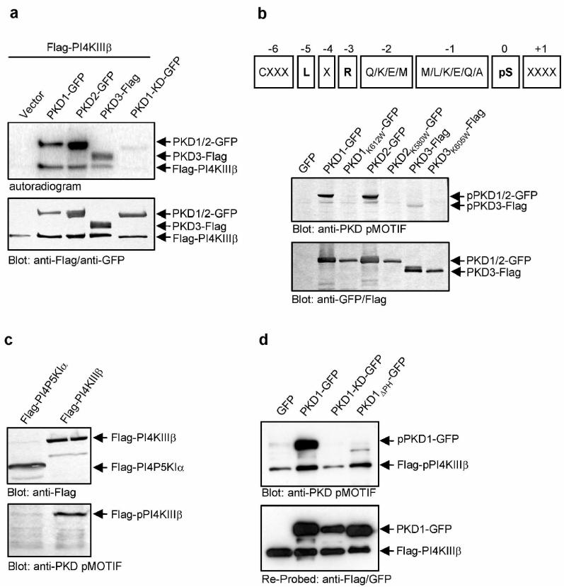
Figure 2.
PKD phosphorylates Flag-PI4KIIIβ in vitro and in whole cells.(a) PKD-GFP phosphorylates Flag-PI4KIIIβ in vitro. HEK293 cells were transfected with plasmids encoding the indicated proteins, lysed and PKD-GFP and Flag-PI4KIIIβ were precipitated using anti-GFP and anti-Flag antibodies. Samples were further processed as described in Methods. (b) A PKD pMOTIF antibody is specific for PKD consensus phosphorylation sites. Upper panel: The sequence of the peptide used for immunization of rabbits, derived from the PKD consensus motif is shown. X represents any amino acid, pS represents phospho-serine. Lower panel: HEK293 transfected with the indicated plasmids were lysed and samples were subjected to SDS-PAGE. Proteins were detected using anti-Flag/anti-GFP or anti-PKD pMOTIF antibodies. (c) The PKD pMOTIF antibody specifically detects PI4KIIIβ. HEK293 transfected with the indicated plasmids were lysed and samples were subjected to SDS-PAGE. Proteins were detected using anti-Flag or anti-PKD pMOTIF antibodies. (d) Expression of PKD1-KD-GFP abrogates detection of Flag-PI4KIIIβ with the anti-PKD pMOTIF antibody. HEK293 cells were transfected with the indicated plasmids. Expression of PKD1-GFP and Flag-PI4KIIIβ proteins was controlled by Western Blot using anti-GFP, anti-Flag and anti-PKD pMOTIF antibodies. Results shown are representative of at least three independent experiments.

