
Fig. 2.
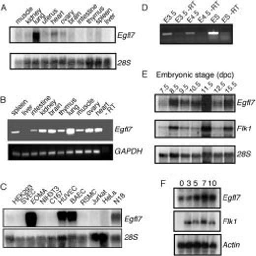
Analysis of Egfl7 expression in adult tissues, cell lines, and during mouse embryogenesis. Northern blots were hybridized with a riboprobe transcribed from the 3′ end of Egfl7. As a loading control, blots were stripped and hybridized with either a 28S riboprobe or an actin riboprobe as labeled. A: Adult mouse tissue expression of Egfl7. B: Reverse transcriptase-polymerase chain reaction (RT-PCR) amplification using the same total RNA from (A) with specific primers to Egfl7 or a loading control, GAPDH. An RT-PCR reaction without the RT is shown in the rightmost lane. C: Egfl7 expression in various mammalian cell lines. D: RT-PCR analysis of Egfl7 expression in embryonic day (E) 3.5 preimplantation and E4.5 peri-implantation mouse blastocysts and in undifferentiated embryonic stem (ES) cells. As a control, duplicate samples of total RNA were amplified without the addition of reverse transcriptase (-RT). E: Egfl7 expression during embryogenesis from E7.5 to E15.5. After hybridization to an Egfl7-specific riboprobe, the blot was stripped and probed with a flk1 riboprobe. F: Total RNA extracted during embryoid body differentiation at 0-, 3-, 5-, 7-, and 10-day time points after differentiation.