
Fig. 4.
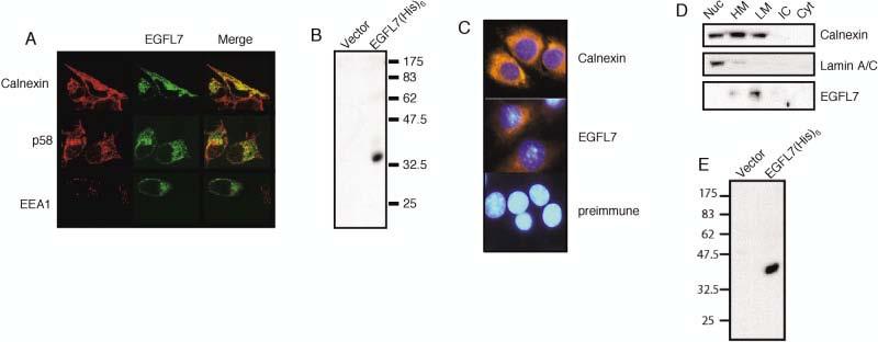
Subcellular localization of EGFL7 in HEK293 and EOMA cells. A: HEK293 cells were stably transfected with an EGFL7–EGFP fusion construct, followed by staining with either Calnexin (endoplasmic reticulum) or p58 (Golgi) specific antibodies. The double-labeled cells were analyzed by confocal microscopy. The rightmost column is an overlay of the green (enhanced green fluorescent protein [EGFP]) and red (Calnexin or p58) channels showing the yellow regions of signal overlap. B: An EGFL7 polyclonal antibody recognizes a single band in transiently transfected HEK293 cells. HEK293 cells were transiently transfected with either empty vector or EGFL7-(His)6 under the control of the CMV promoter, followed by Western blot analysis using an affinity purified polyclonal EGFL7 antibody. C: Indirect immunofluorescence analysis of Calnexin and EGFL7 in EOMA cells. Nuclear staining was achieved by staining with Hoechst 33342. D: HEK293 cells were stably transfected with an EGFL7–(His)6 fusion and fractionated by differential centrifugation. Protein samples from each cellular fraction were separated in 12% sodium dodecyl sulfate-polyacrylamide gel electrophoresis and analyzed by Western blot with EGFL7, Calnexin, or Lamin A/C specific antibodies. Nuc, nuclear; HM, heavy microsome; LM, light microsome; IC, insoluble cytoplasmic; Cyt, soluble cytoplasmic. E: HEK293 EBNA1 cells were transiently transfected with an EGFL7–(His)6 fusion construct under the control of the CMV promoter. Samples from the media of mock-transfected and EGFL7–(His)6-transfected cells were harvested 2 days post-transfection and analyzed by Western blot using an HRP-conjugated anti-(His)6 antibody.