
Fig. 5.
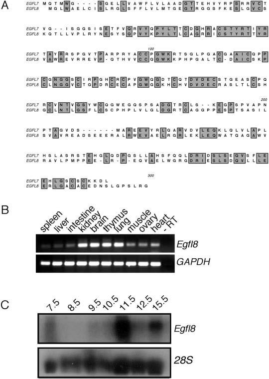
Cloning and expression of the EGFL7 mouse paralog EGFL8. A: Alignment of the mouse EGFL7 and EGFL8 proteins. Regions of identity are indicated by grey shading. B: Reverse transcriptase (RT)-polymerase chain reaction analysis of Egfl7 and Egfl8 expression in adult mouse tissues. Specific intron-spanning primers that amplify Egfl7, Egfl8, or GAPDH were used for polymerase chain reaction amplification. C: Northern blot analysis of Egfl8 expression during embryogenesis from embryonic day (E) 7.5 to E15.5. As a loading control, blots were stripped and hybridized to an 28S riboprobe.