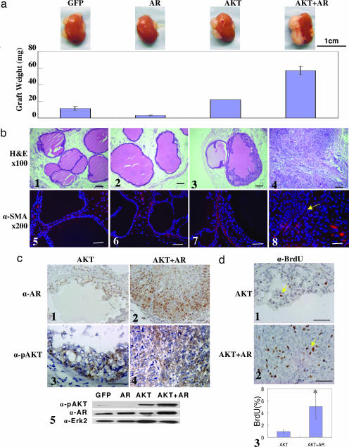
Fig. 1.
AKT and AR synergize and progress naïve prostatic epithelium to invasive adenocarcinoma in a dissociated prostate cell regeneration method within 6–8 weeks. (a) Weight (± SEM) of regenerated tissues derived from prostatic epithelial cells infected by the indicated lentivirus combinations (n ≥ 4). The micrograph above each bar shows a representative picture of the regenerated tissues attached to the murine kidneys. (Scale bar: 1 cm.) (b) H&E staining of the regenerated tissues (b1–b4) and IHC analysis for SMA in the regenerated tissues (b5–b8). The yellow arrow indicates the absence of the SMA-positive stromal layer. (c) IHC staining for AR (c1 and c2) and phospho-AKT (c3 and c4) in regenerated tissues in the AKT and AKT-plus-AR groups, respectively, and Western blot analysis of the AR and phospho-AKT (c5). Erk2 is used as a loading control. (d) IHC analysis for BrdU in regenerated tissues in the AKT (d1) and AKT-plus-AR (d2) groups. (d3) Quantification of BrdU-positive cells in the AKT and AKT-plus-AR groups (∗, P < 0.001). (Original magnification: ×200. Scale bars: 100 μm.)

