
FIGURE 6.
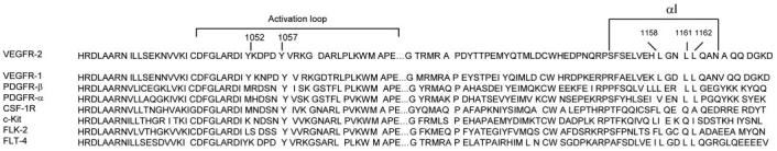
Structure-based amino acid sequence alignment of the kinase domain of VEGFR-2 with the other type III RTKs. The secondary structure of VEGFR-2 indicates that the COOH-terminal lobe of the kinase domain is folded into 7 α-helices and 2 β-sheets (23). Residues from 1150 to 1164 constitute αI. The αI is the last helix and it separates the kinase domain from the carboxyl terminus of VEGFR-2. The position of activation loop tyrosine autophosphorylation sites Tyr-1052 and Tyr-1057 are also shown. Partial sequence alignment of the kinase domain and activation loop regions of human VEGFR-1 and various receptor tyrosine kinases of type III family are shown.