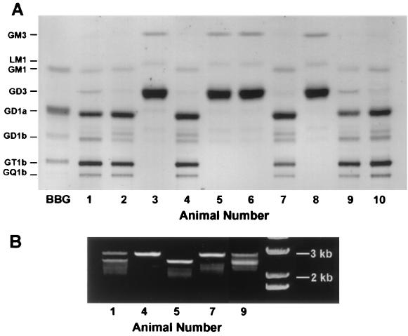
Figure 1.
Ganglioside pattern and genotype analysis of one representative litter. (A) Thin-layer chromatogram of gangliosides purified from the cerebra of 10 littermate pups. Isolated material corresponding to 0.8 mg protein was applied at the origin, and the plate developed in chloroform-methanol-0.25% aqueous KCl (5:4:1); ganglioside bands were revealed with resorcinol spray. Pups nos. 3, 5, 6, and 8 were deficient in complex gangliosides and showed an excess of GM3 and LM1 together with a very large accumulation of GD3. Pups nos. 1 and 9 were heterozygotes (see below) and contained complex gangliosides similar to wild type (nos. 2, 4, 7, and 10) with modest elevation of GD3 accompanied by reduction of a ganglioside running just ahead of GD1b. BBG, bovine brain gangliosides (standards). (B) Genotype identification. DNA isolated from the above brain tissues was subjected to PCR and the resulting products developed in a 0.7% agarose gel. The 2.9- and 2.5-kb bands represent wild-type and mutant GalNAc-T alleles, respectively. Pups nos. 4 and 7 were identified as wild type, nos. 1 and 9 as heterozygotes, and no. 5 as knockout. The far right column is DNA ladder standards (GIBCO).

