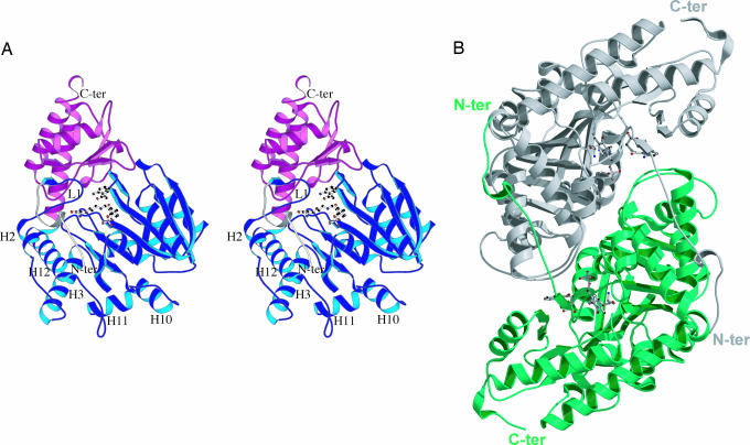
Fig. 1.
Ag-HKT molecular architecture. (A) Stereo ribbon representation of the Ag-HKT subunit. The N-terminal extension, the large domain, and the small domain are colored in white, blue, and magenta, respectively. The loop protruding into the active site and carrying the cisPro-24-Gly-25 motif is labeled L1. The deep cleft hosting the enzyme active site is placed at the domain interface, where the PLP cofactor and the inhibitor molecules are shown in ball-and-stick representation. The N and the C termini are indicated, as are the helices connected by the loops involved in dimer stabilization. (B) Ribbon representation of the functional Ag-HKT homodimer seen along the dyad axis. The two subunits are colored white and green; PLP and inhibitor molecules are drawn in ball-and-stick representation. The N and the C termini are indicated. The figure was generated with the program molscript (50).

