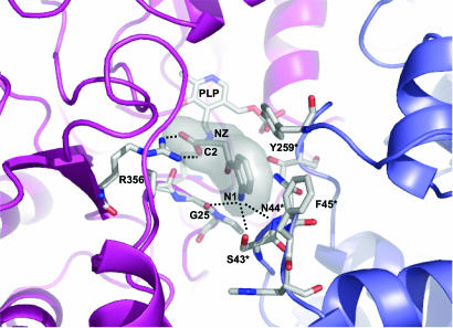
Fig. 3.
Closeup view of the enzyme active site in the Ag-HKT:INI structure. The protein portions building up the active site and contributed by the two subunits of the functional homodimer are shown in a ribbon representation and colored blue and magenta. The PLP cofactor, the inhibitor molecule, and the protein residues involved in ligand binding are shown. The inhibitor C2 atom equivalent to the Cα of the physiological amino acid substrate is indicated. The major interactions established between the inhibitor and residues of Ag-HKT are indicated as dotted lines. The figure was generated by using pymol (www.pymol.org).

