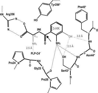
Scheme 2.
Schematic view of the Ag-HKT active site. The key interactions involved in inhibitor binding are indicated together with the distances observed in the Ag-HKT:INI structure. The chemical groups featuring the physiological substrate 3-HK are shown in light gray, and their interactions with the protein residues, resulting from manual modeling, are indicated. The arrow indicates the C4 position on the 4-(2-aminophenyl)-4-oxobutyric acid that would carry the proposed chemical modification consisting of the replacement of the carbonyl group by an oxy-amino (-O-NH2) moiety.

