Fig. 2.
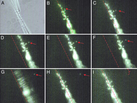
Successive frames of a pulling operation. The red arrow indicates the position of a single cell being pulled. (A) Phase contrast showing the tip of a suction pipette holding a C. crescentus cell in place, which is to be pulled from the flexible thin pipette. (B–I) Fluorescence images using NanoOrange to label the cell body, stalk, and holdfast. (C–F) The cell is pulled, and the thin flexible pipette increasingly deviates from its original position (indicated by the red dotted line). (F) The instant right before the cell is pulled off from the thin flexible pipette. (G) The cell is successfully pulled off the thin flexible pipette; the movement of the pipette is easily seen as a blurring of the attached cells. (H) The thin flexible pipette has returned to its original position. (I) The red bracket outlines the cell stalk taken inside the suction pipette following the cell body, a moment after their detachment from the substrate. (Magnification: ×500.) See Movie 1 for a demonstration of the pulling experiment.

