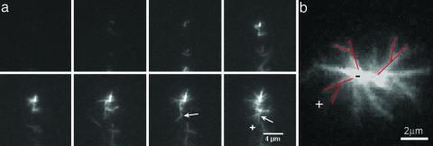
Fig. 2.
Total internal reflection fluorescent images of an aster formation. Conditions were 1 μM actin monomer, 200 nM GST-VCA, and 10 nM Arp2/3 complex. (a) Images in the time-lapse series are spaced 30 s apart for a total time of 3.5 min. The experiments begin with buffer alone in the chamber. Actin, Arp2/3 complex, and activator are quickly mixed and pulled through the chamber. High concentrations of Arp2/3 complex lead to the formation of asters. Most likely, very small spontaneously nucleated seeds act as nucleation points from which Arp2/3 complex rapidly forms branches autocatalytically. The initial seeds are smaller than the optical resolution and thus appear as points at the center of the aster. Formation of a new branch (≈70° is characteristic to Arp2/3 branching) marked by a white arrow in the two last frames shows that the actin filaments barbed ends (+) point outward. (b) An image of the aster part that is close to the coverslip surface clearly shows branches pointing away from the aster center, three of them are marked in red. The growth direction is thus from the pointed (−) end to the barbed (+) end.

