Fig. 1.
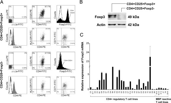
Expression of Foxp3 mRNA in CD4+ regulatory T cell lines derived from MS patients immunized with irradiated autologous MBP-reactive T cells. (A) Two representative patterns of CD4+ regulatory T cell lines by flow cytometry. Representative T cell lines (lines B9 and F8) were analyzed for the expression of CD4 paired with that of CD25, HLA-DR, CD71, and Foxp3. Open curves in the histograms represent isotype control. Staining with specific antibody to CD4 or Foxp3 is indicated by gray or solid curves, respectively. (B) Immunoblot analysis of the same T cell lines for protein expression of transcription factor Foxp3. (C) Foxp3 mRNA expression by real-time PCR in all 30 CD4+ regulatory T cell lines and six reference MBP-reactive T cell clones derived from the same patients.

