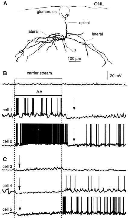
Figure 1.
Odor-evoked responses in mitral cells. (A) Projection of the reconstruction of the morphology of a mitral cell recorded in vivo with apical and lateral dendrites. The outline of a glomerulus is shown schematically. (B) Examples of odor-evoked excitatory responses. AA but not the carrier stream (cell 1) evokes APs (up to 250 s−1) in mitral cells (APs are clipped). Moderate or high odor-evoked firing rates result in abrupt decreases in firing rates and hyperpolarizations (arrows) following termination of the odor presentation. (C) Examples of odor-evoked hyperpolarizations (arrows). AA under control conditions caused an initial hyperpolarization, followed by a transient burst of spikes after termination of the stimulus. Odor presentations are 5 s.

