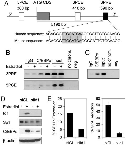
Fig. 4.
Id1 is a direct target gene of C/EBPα and important for myeloid differentiation. (A) Genomic locus of the Id1 gene. The gray box represents the coding sequence including one intron, and the white and black boxes represent elements conserved between mouse and human. Within the black box C/EBPs have been shown to bind to an 8-bp sequence (highlighted in gray) (19). 5PCE, 5 prime conserved element; ATG CDS, ATG coding sequence; 3PCE, 3 prime conserved element. (B) ChIP analysis of K562αER cells treated with vehicle alone or with 1 μM β-estradiol for 24 h. Note the increased signal for C/EBPα binding to the 3PRE after induction. The PCR product observed in cells without C/EBPα induction probably is caused by spontaneous translocation of a small amount of C/EBPα-ER in the absence of β-estradiol. This hypothesis is supported by the observation that no binding was detected in parental K562 cells (data not shown). No binding to the 5 prime conserved element (5PCE) was detected. (C) ChIP analysis of mouse BM demonstrated C/EBPα binding to the 3PRE in vivo. no chrom., no chromatin; neg., negative. (D) Western blot of K562αER cells expressing short hairpin RNA (shRNA) against Id1 (siId1) or a control short hairpin RNA (siGL). Cells were treated with 20 nM β-estradiol. This concentration was used, because up-regulation of C/EBPε and Id1 induction was comparable to treatment with 1 μM β-estradiol, but the cells were more viable. Id1 induction upon C/EBPα activation was suppressed and accompanied by severely impaired C/EBPε induction. (E) Flow cytometry showed decreased expression of CD11b (P < 0.05) and impaired down-regulation of glycophorin A (GPA) (P < 0.05) upon suppression of Id1 after 24 h. For this analysis, cells with a high GFP expression were gated.

