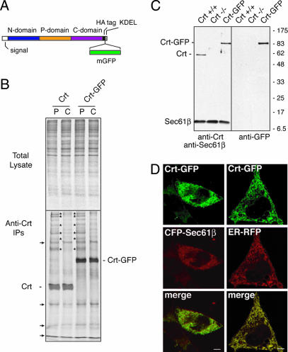
Fig. 1.
Crt-GFP construction and characterization. (A) Illustration of functional domains of Crt and the insertion sites for an hemagglutinin (HA) epitope tag and monomerized GFP. (B) crt−/− cells were transiently transfected with either Crt or Crt-GFP and analyzed 40 h later by pulse–chase labeling and native immunoprecipitation. Pulse labeling (P) with [35S]methionine was for 30 min, followed by chase (C) in unlabeled media for 90 min. Total cell lysates from the samples (Upper) and the anti-Crt immunoprecipitates (Lower) are shown. The positions of Crt and Crt-GFP are indicated. Several coprecipitating proteins that transiently interact with both Crt and Crt-GFP during the pulse and release during the chase period are indicated by asterisks. Background bands that were also seen in control transfected cells (see Fig. 6) are indicated by the arrows. (C) Immunoblots of whole cell lysates from crt+/+, crt−/−, or Crt-GFP cells (crt−/− cells stably expressing Crt-GFP). (Left) Probed with anti-Crt and anti-Sec61β (a loading control). (Right) The same blot stripped and reprobed with anti-GFP. (D) Complete colocalization of Crt-GFP in live cells with cotransfected ER markers: mCFP-Sec61β (Left) or ER-RFP (Right). (Scale bars, 5 μm.)

