Fig. 3.
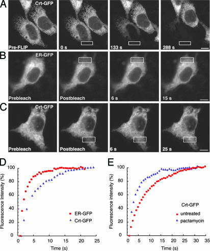
Dynamics of Crt-GFP. (A) Images of a cell expressing Crt-GFP before (Left) and at various times during repeated photobleaching in the region outlined by the white box. Fluorescence in the bleached cell was depleted uniformly over time. (B and C) FRAP analysis of quiescent cells (treated with Pact for 1 h; Fig. 2A) expressing ER-GFP or Crt-GFP. Images were captured immediately before (Prebleach), immediately after (Postbleach), and at times after photobleaching in the area outlined by the white box. Both ER-GFP and Crt-GFP are highly mobile, because unbleached fluorescent proteins rapidly diffuse into the bleached regions. [Scale bars, 5 μm (A–C).] (D) Plots of recovery rates reveal that ER-GFP (red squares) diffuses more rapidly than Crt-GFP (blue triangles). (E) FRAP analysis of Crt-GFP in cells actively translating new proteins (untreated, red squares) or under quiescent conditions (Pact-treated, blue triangles) demonstrates that Crt-GFP mobility decreases in the presence of substrates.

