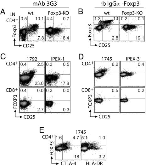
Fig. 1.
Flow cytometric detection of Foxp3 in murine and human cells. (A and B) Normal or Foxp3-deficient mouse lymph node cells were stained for Foxp3 and cell-surface markers by using digoxigenin-conjugated mAb 3G3 (A) or Foxp3-specific rabbit antibody (B). CD4+ gated lymphocytes are shown. (C–E) Normal (1792 and 1745) or FOXP3-deficient (IPEX) PBMC were stained for FOXP3 and lymphocyte markers by using digoxigenin-conjugated mAb 3G3 (C) or digoxigenin-conjugated Foxp3-specific rabbit antibody (D and E). Both CD4+ and CD8+ gated lymphocytes are shown. Additional IPEX-1 PBMC were not available for subsequent analysis with rabbit antibody. High background staining of Foxp3− cells is a consequence of the three-step staining procedure.

