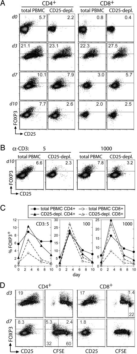
Fig. 2.
Analysis of FOXP3 expression in activated human CD4+ and CD8+ T cells. (A–C) Total or CD25-depleted PBMC from donor 1745 were stimulated with 5, 100, or 1000 ng/ml anti-CD3. FOXP3 and CD25 expression on CD4+ and CD8+ cells were assessed at days 3, 7, and 10 of culture. Shown are expression profiles for 100 ng/ml anti-CD3 (A), 5 or 1,000 ng/ml anti-CD3 for CD4+ gated cells (B), and the plotted percentage of gated cells expressing FOXP3 (C). FOXP3 was detected with digoxigenin-conjugated FOXP3-specific rabbit antibody. (D) Total PBMC from donor 1745 were labeled with CFSE and stimulated with 100 ng/ml anti-CD3. FOXP3 expression was assessed at days 3 and 7 with digoxigenin-conjugated rabbit antibody. Data are representative of four separate experiments and three normal adult donors.

