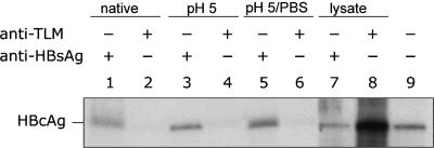
Fig. 3.
Cleavage of HBsAg by endosomal proteases results in surface exposure of the TLM. Purified HBV particles were subjected to immunoprecipitation with either an HBV-TLM-specific antiserum (lanes 2, 4, 6, and 8), or an HBsAg-specific serum as a positive control (lanes 1, 3, 5, and 7). The precipitated material was immunoblotted by using an HBcAg-specific antiserum. In lanes 1 and 2 precipitates of untreated HBV particles were loaded using an HBsAg-specific serum or a TLM-specific serum. Purified HBV particles were incubated for 30 min at pH 5.0 and immunoprecipitated by using an HBV-TLM-specific antiserum (lane 4) and an HBsAg-specific serum (lane 3). Purified HBV particles were incubated for 30 min at pH 5.0, then by addition of 10× PBS the pH was shifted to ≈7. Afterward, immunoprecipitation was performed by using an HBV-TLM-specific antiserum (lane 6) and an HBsAg-specific serum (lane 5). Purified HBV particles were incubated for 30 min at pH 5.0 in endosomal lysate from HepG2 cells and immunoprecipitated by using an HBV-TLM-specific antiserum (lane 8) and an HBsAg-specific serum (lane 7). Recombinant HBcAg (lane 9) served as positive control.

