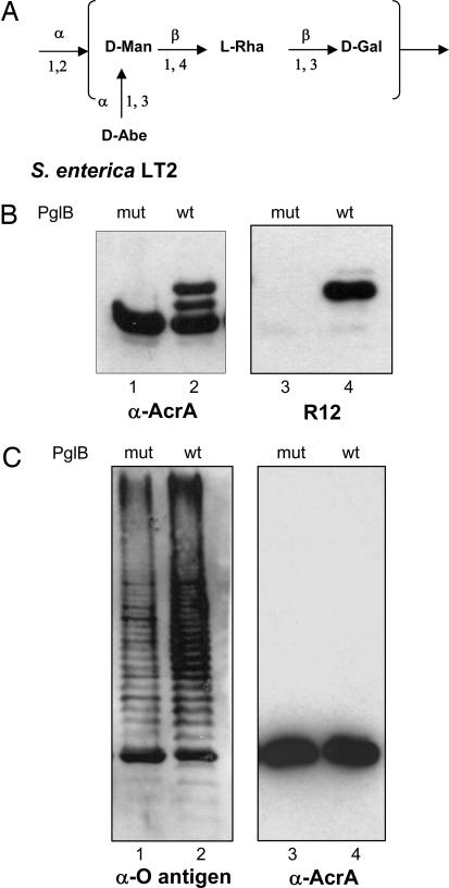
Fig. 3.
Glycosylation of AcrA in S. enterica LT2. (A) Structure of the Salmonella LT2 O-antigen subunit. Man, mannose; Rha, rhamnose; Abe, abequose. (B) Western blot analysis of S. enterica SL3749 (ΔWaaL) periplasmic extracts expressing AcrA, and the pgl locus containing either PglBwt or PglBmut. Antisera anti-AcrA (lanes 1 and 2) or R12 (lanes 3 and 4) were used. (C Left) Western blot analysis of S. enterica SL3749 whole-cell extracts expressing AcrA and PglBwt or PglBmut in the absence of the pgl locus, showing production of S. enterica LT2 Und-PP-bound O antigen. Crossreaction of the antisera with AcrA can be detected. (C Right) Western blot of purified AcrA from periplasmic extracts of the cells is shown. Only one band, corresponding to the unglycosylated protein, is detectable in the presence of PglBwt or PglBmut.

