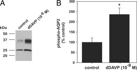
Fig. 3.
Quantification of AQP2 phosphorylation in response to short-term dDAVP treatment by immunoblotting. (A) IMCD suspensions treated with dDAVP (10−9 M) or without (control) for 10 min. Immunoblots were probed by using a phosphospecific AQP2 antibody that recognizes phosphorylated S256. (B) Phosphorylated AQP2 levels were significantly increased with dDAVP treatment (dDAVP 237 ± 31.4% vs. control 100 ± 21.5%; ∗, P < 0.05).

