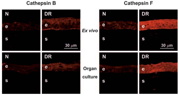
FIGURE 3.

Distribution of cathepsins B and F in ex vivo and organ-cultured normal (N) and DR corneas. Note a slight increase of cathepsin B (left panels, polyclonal antibody [pAb] N-19) and a significant increase of cathepsin F (right panels, pAb V-20) in DR corneal epithelium. Data for ex vivo and organ-cultured corneas are similar. Indirect immunofluorescence. e, epithelium; s, stroma.
