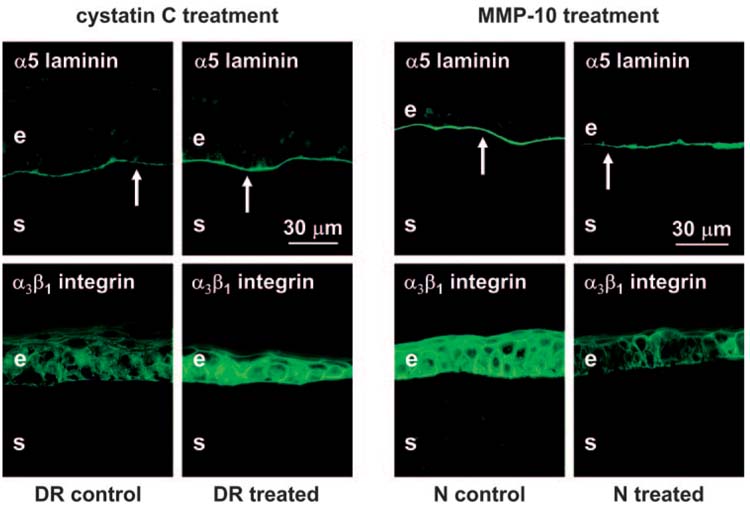
FIGURE 6.

Left panels: Treatment of wounded DR organ-cultured corneas with cathepsin inhibitor cystatin C. Healed corneas are presented. Laminin-10 α5 chain staining (arrows) becomes continuous and similar to normal after cystatin C treatment (upper panels). Integrin α3β1 staining is weak and irregular in control corneas but becomes significantly stronger and more regular in cystatin C-treated corneas (lower panels). Right panels: Treatment of wounded normal organ-cultured corneas with MMP-10. Healed corneas are presented. Note continuous staining of the epithelial BM (arrows) for laminin-10 α5 chain in BSA-treated (control) corneas and discontinuous staining in MMP-10-treated corneas (upper panels). Strong staining for integrin α3β1 is seen in control corneas; it is weak and irregular in treated corneas (lower panels). Indirect immunofluorescence. e, epithelium; s, stroma.
