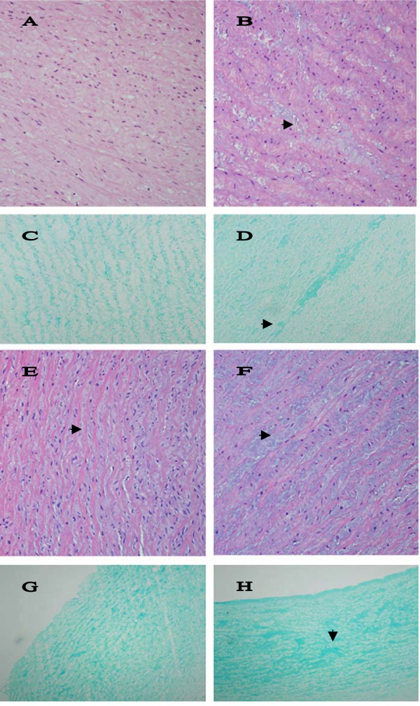
Figure 7.
Histopathological changes in arteries. A. The media of normal aortic arch shows orderly aligned elastic fibers and very little PGs between them. Hematoxylin & eosin, magnification × 200. B. Small pools of PGs (▶) separate fibers and cells in the media of DSLD-affected aortic arch. Hematoxylin & eosin, magnification × 200. C. Alcian blue stains material aligned closely with elastic fibers in normal arch, magnification × 200. D. Pools of PGs (▶) stain strongly with alcian blue in the media of DSLD-affected arch, magnification × 200. E. The media of normal coronary artery shows orderly aligned elastic fibers separated by thin layers of PGs (▶) between them. Hematoxylin & eosin, magnification × 200 (× 200). F. Small pools of PGs (▶) separate fibers and cells in the media of DSLD-affected coronary artery. Hematoxylin & eosin, magnification × 200. G. Alcian blue stains material aligned closely with elastic fibers in the media of normal coronary artery, magnification × 100. H. Pools of PGs (▶) stain strongly with alcian blue in the media of DSLD-affected coronary artery, magnification × 100.

