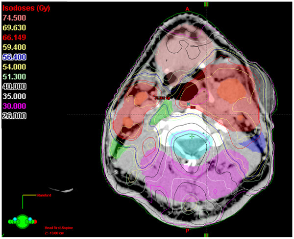
Figure 6.
An example of an IMRT isodose plan using simultaneously integrated boost. Depicted is an axial slice, 64 mm above the isocenter of the plan. Contoured are PTV1 (69.6 Gy), PTV2 (60 Gy) and PTV3 (54 Gy), gross tumor volumes of the primary and macroscopic nodal disease, and normal structures (spinal cord, brain, parotid glands, anterior soft tissues, dorsal soft tissues). Note the well-spared spinal cord and parotid glands despite of bilateral nodal disease covered with high doses (nodal and primary gross tumor volumes included into the PTV1).

