
Fig. 1.
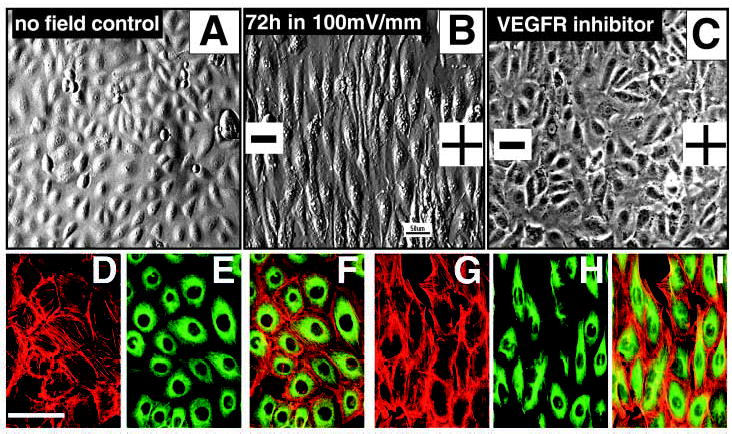
Perpendicular orientation and elongation of endothelial cells in a small physiological EF. (A) Control HUVEC cells cultured in the same chamber without EFs showed a typical cobblestone morphology and random orientation. (B) Cells exposed to small applied EFs showed dramatic elongation and perpendicular orientation in the EF. (C) Cells treated with a VEGFR inhibitor that completely abolished perpendicular orientation and significantly inhibited elongation in an applied EF (72 hours, 100 mV mm−1). (G-I) Most actin filaments (red) and microtubules (green) became aligned along the long axis of the cells (12 hours at 150 mV mm−1). (D-F) No-field controls showed no obvious alignment and cell elongation. (A,B) Images taken with Hoffman modulation optics. (C) Image taken with phase-contrast optics. (F,I) Merged images.