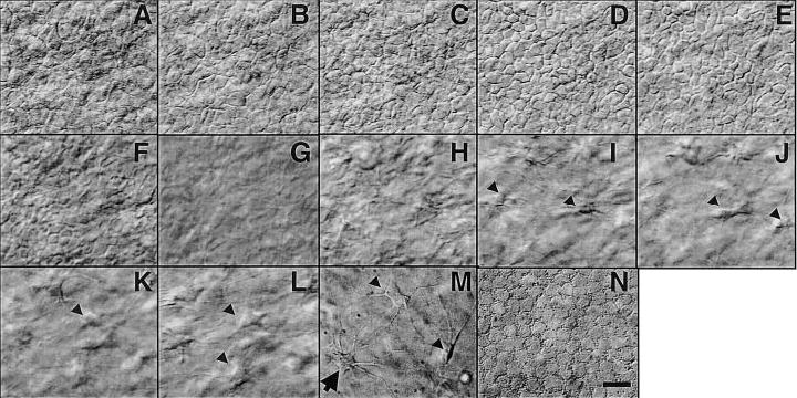
Figure 1.

Hoffman optical sections through whole excised live bovine cornea. Individual epithelial cells (mosaic-like structures in panels A–E), an acellular layer (G), fibro-blasts in the stroma (arrow in panels H–M), and a possible dendritic cell (arrow in panel M), and endothelial cells (N) can be seen directly. Bar = 10 μm. Video 01 at http://www.fasebj.org/cgi/content/full/17/3/397/F1/DC1.
