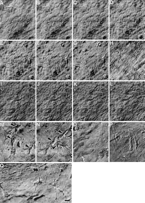
Figure 2.

Hoffman optical sections through whole excised live rat (A–H) and human (I–Q) cornea. Live epithelial cells (mosaic-like structures in panels A–E, I–K), fibroblasts in the stroma (arrow in panels M, N, P, Q), and endothelial cells (H, O) can be seen directly. In cornea from old donors, webs of fibrous structure can be seen (P, Q). Bar = 10 μm. Video 02 at http://www.fasebj.org/cgi/content/full/17/3/397/F2/DC1.
