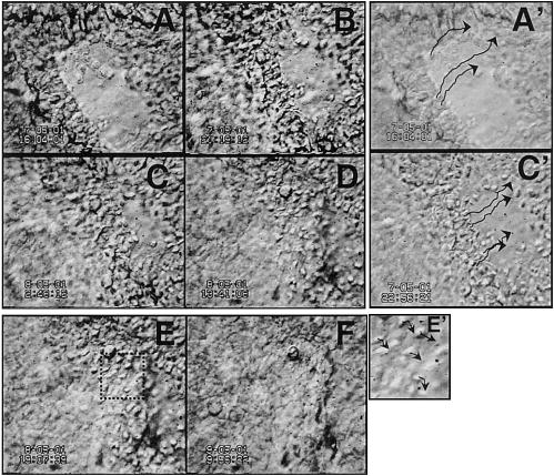
Figure 4.

Dynamics of cell behavior during wound healing in situ can be observed directly. Stratified corneal epithelium healing in whole organ culture. Epithelial movements during healing (A–D), wound fronts meet (D), epithelial reconstruction (E, F). A′, C′, E′) show paths of single cells. Arrows indicate trajectories of individual cells tracked by computer. Video 04, http://www.fasebj.org/cgi/content/full/17/3/397/F4/DC1. Note: these images were recorded with low magnification, thus low resolution, to show healing of a whole wound.
