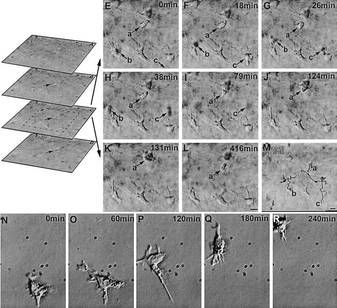
Figure 7.

Cell movements in the stroma. Spatial sequence (A–D, optical sections of the stroma) shows the focal planes above (A, B) and below (D) focal plane C (video 06). Time sequence of optical section C (E–L) and trajectories of migratory cells superimposed with the focal plane C (M) (video 07). Migratory cells (presumably dendritic cells, arrow a, b, c) and the rest spiny stationary cells can be readily distinguished. Note that the migratory cells b, c are moving in and out of the focal plane, therefore they can appear as dark shadows, while cell a remains in the focal plane throughout the sequence, moves toward and contacts a keratinocyte (K), then moves away (L). Movie and M shows their paths. Cell b moves into the field at 18 min, then rapidly moves out of the focal plane, with a shadow visible at 26 and 38 min frames and fades away in 79 min frame. The keratinocytes remained static and did not show any obvious movement during the recording period. See online movie, video 08 showing A–D, video 09 showing E–L). N–R) A dendritic cell in isolated culture moves with similar morphology, but 10-fold faster. Supplemental video 08 at http://www.fasebj.org/cgi/content/full/17/3/397/F7/DC1; video 09 at http://www.fasebj.org/cgi/content/full/17/3/397/F7/DC2; video 10 at http://www.fasebj.org/cgi/content/full/17/3/397/F7/DC3.
