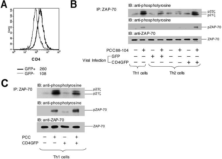
Figure 5.

Th1-like pattern of proximal TCR-induced protein tyrosine phosphorylation in Th2 cells with restored CD4 expression. (A) Polarized Th1 and Th2 5C.C7 T cells were infected with recombinant retrovirus encoding either EGFP or a mouse CD4-EGFP fusion protein. The infected cells were then subjected to cell sorting for EGFP-positive cells. Infected cells expressing CD4-EGFP showed restoration of CD4 expression to a level typical of polarized Th1 cells, which is ∼2.5-fold greater than that of the uninfected EGFP-negative Th2 cells from the same culture. (B) Th1 cells, EGFP-negative Th2 cells, and Th2 cells expressing either EGFP or CD4-EGFP were stimulated with antigen-presenting cells pulsed with 100 μM PCC88–104 peptide. Five minutes later, the cells were lysed, and the lysates were subjected to immunoprecipitation and immunoblotting analysis as described in Fig. 2. Results are representative of three experiments. (C) Polarized Th1 cells were either left uninfected or infected with retrovirus encoding CD4-EGFP and sorted for EGFP expression. These cells were then stimulated with antigen-presenting cells pulsed with 100 μM PCC88–104 peptide, lysed, and the lysates subjected to immunoprecipitation with anti-ZAP-70 antibody followed by SDS-PAGE analysis and immunoblotting with either anti-phosphotyrosine or anti-ZAP-70 antibody.
