
Figure 3.
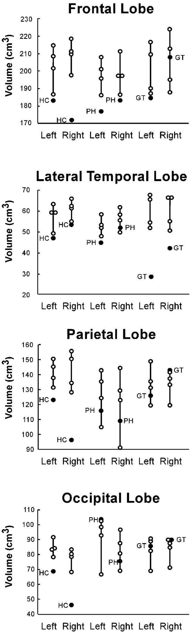
Volumes of Major Brain Regions for Three Amnesic Patients with Large Medial Temporal Lobe Lesions and Additional Damage to Neocortex
Patients are represented by filled circles. Separate control groups (n = 4; unfilled circles) were matched to patient H.C., patient P.H., and patient G.T.