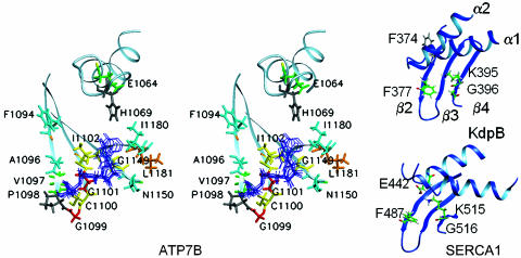
Fig. 6.
Organization of the nucleotide-binding site in the N-domains of ATP7B, SERCA1, and KdpB. (A) Stereo view of the ensemble of the ATP molecules docked into the ATP-binding site of ATP7B. The residues in proximity to ATP are colored according to the magnitude of the ATP-dependent secondary chemical shifts as in Fig. 4A. Data are not available for H1069 (shown in gray). (B) Fragments of the KdpB and SERCA1 N-domain structures with the residues involved in ATP binding.

