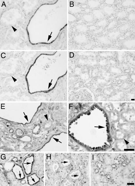
Fig. 2.
Phospho-mTOR, -S6K, and -S6 are specifically induced in cyst-lining epithelial cells in ADPKD and mouse models. (A–D) Renal tissue sections isolated from human ADPKD patients (A and C) or normal patients (B and D) were analyzed by immunohistochemistry using antibodies against phospho-mTOR (Ser 2448) (A and B) or phospho-S6K (Thr 389) (C and D). Note specific staining of cystic epithelial cells (A and C, arrows) but not surrounding normal epithelium (A and C, arrowheads). (E–I) Renal tissue sections derived from Pkd1cond/cond:TgN(balancer1)2Cgn (E) and MAL-overexpressing (F) mice, nontreated (G) or rapamycin-treated (H) (5 mg/kg of body weight) oprk-rescue (Tg737orpk/orpk;TgRsq) mutant mice, and nontreated orpk-heterozygous-rescue (Tg737orpk/+;TgRsq) (I) control mice were immunostained by using an antibody against phospho-S6 ribosomal protein (Ser 235/236). Note specific cytoplasmic staining of cystic-epithelial cells (E–G, arrows) but not surrounding normal epithelium (E and F, arrowheads; and I). The cyst-specific cytoplasmic immunosignal is abolished after rapamycin treatment (H, arrows). (Scale bars, 20 μm.)

