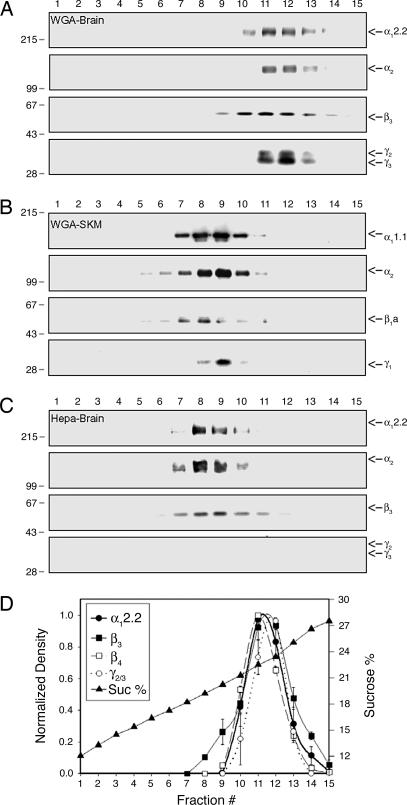
Fig. 1.
Comparison of the Ca2+ channel complex partially purified from different tissues or through different methods. Sucrose gradient fractionation of Ca2+ channel complexes and subsequent immunoblotting for Ca2+ channel subunits demonstrate that the size of partially purified Ca2+ channel complexes differs depending on tissue and purification method. (A) Sucrose gradient fractionation of WGA-bound Ca2+ channel complex from rabbit brains. (B) Sucrose gradient fractionation of WGA-bound Ca2+ channel complex from rabbit skeletal muscles (SKM). (C) Sucrose gradient fractionation of heparin-bound Ca2+ channel complex from rabbit brains. The numbers at the top indicate the fraction of the sucrose gradient from top to bottom. Molecular mass standards (×10−3) are indicated on the left side of the panels. (D) Densitometry of Ca2+ channel subunits from Western blots of sucrose gradient fractions of WGA-bound Ca2+ channel complex. Fraction #, fraction number of sucrose gradient; Sucrose %, percentage of sucrose.

