
FIGURE 2. Matrigel invasion of IOMM-Lee cells transfected with mock, EV/SV, pU, pUR and pU2.
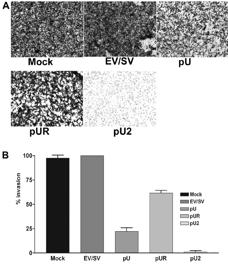
1×10⁶ transfected cells were seeded on matrigel-coated 12-well transwell inserts (8-μm pores) and allowed to invade. After 24 h, cells that had passed through the filter into the lower wells were fixed and stained with Hema 3 and photographed (A). Percentage of invasion was quantitated as described in Materials and Methods (B).