
FIGURE 3. Spheroid migration by IOMM-Lee cells transfected with mock, EV/SV, pU, pUR and pU2.
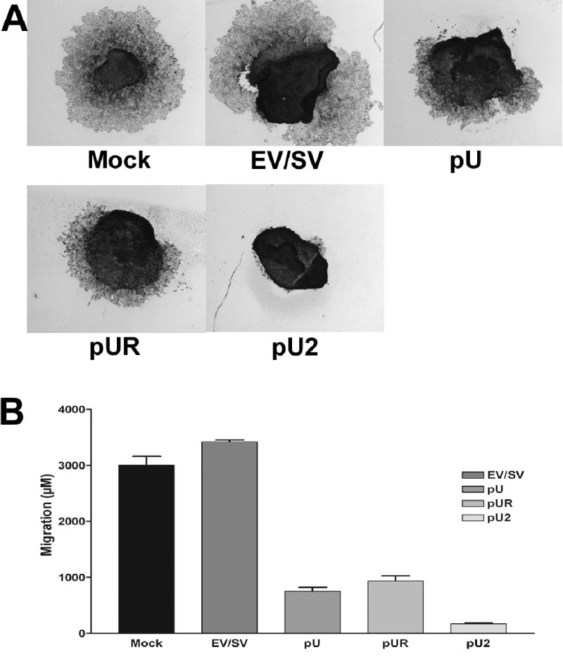
Spheroids of 100–200 microns were prepared by seeding 5×10⁴ cells on 96-well low attachment plates by continuous shaking at 60 rpm. The spheroids were transfected with mock, EV/SV, pU, pUR or pU2. 72 h later, the spheroids were placed on vitronectin-coated 8-well chamber slides and allowed to migrate. After 48 h, the spheroids were then fixed and stained by Hema 3 (A). Spheroid migration was quantified and graphically represented (B).