
FIGURE 5. Regression of subcutaneous tumors by RNAi.
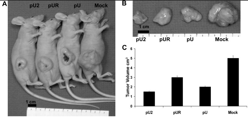
5×10⁶ IOMM-Lee cells were injected subcutaneously into the right flank of nude mice. When the tumors reached 4–5mm in size, pU, pUR or pU2 were injected intra tumorally on alternate days (A). A total of five injections were given. Tumors were excised after 4 weeks and photographed (B) and tumor volume determined (C).