
FIGURE 6. Suppression of intracranial tumor growth by RNAi.
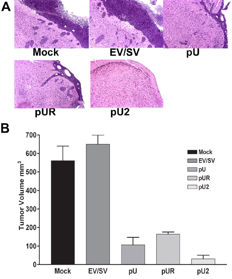
0.5×10⁶ IOMM-Lee parental cells or cells transfected with EV/SV, pU, pUR or pU2 were injected intracranially into nude mice. After 4 weeks, the mice were sacrificed, the brains removed and fixed in formaldehyde, paraffin embedded and sectioned followed by H&E staining (A). The stained sections were viewed under microscope and the tumor area determined in serial sections to approximately determine the intracranial tumor volume (B).